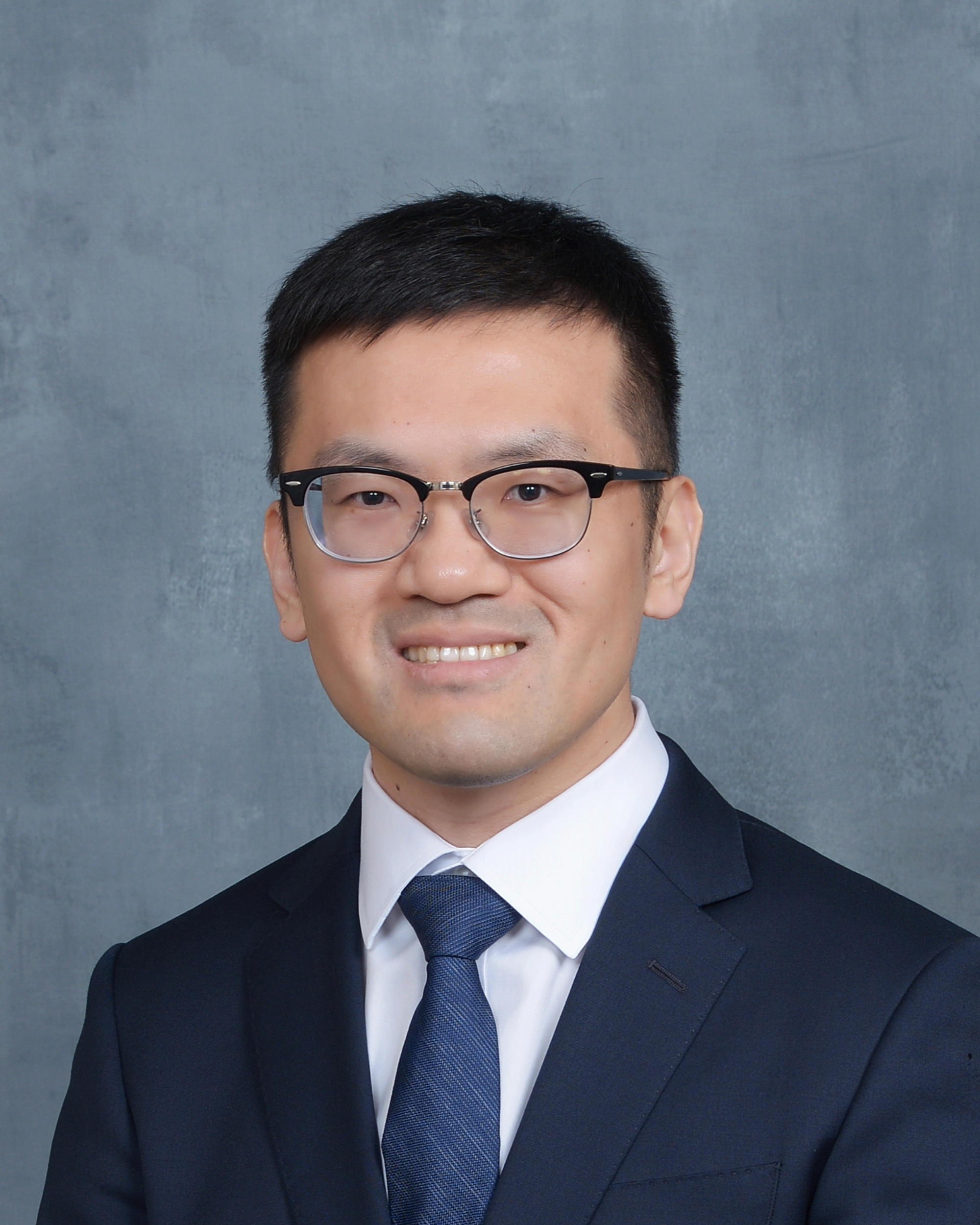
About me
I am an Assistant Professor of Department of Radiology, Wake Forest University School of Medicine. My work focuses on developing artificial intelligence (AI) algorithms for 1) medical image generation and 2) AI-assisted disease diagnosis.
AI especially deep learning (DL) has achieved great success in recent years, ranging from computer vision to natural language processing. The emerging of generative AI models like ChatGPT and Sora, with the ability of recognizing and generating contents, has demonstrated huge potentials to impact human lives in future. In terms of medical imaging, DL has been extensively used in multiple applications such as medical imaging quality improvement, disease diagnosis, and treatment planning. As of October 19, 2023, Food and Drug administration (FDA) has approved nearly 700 AI-enabled devices and projected an over 30% increment in 2023.
AI-based medical image generation will not only result in higher quality medical images, but also contribute to lower medical imaging scan costs. My previous work on MRI super-resolution significantly improves image quality by showing more clear tissue boundaries and texture details comparing with fast MRI scan with poor image quality. For cardiac MRI, my proposed neural network with convolutional long short-term memory greatly improves both spatial and temporal image quality by removing motion artifacts and predicting intermediate time point images. In terms of lowering medical imaging costs, I am working on reducing the amount of contrast medical in contrast-enhanced MRI and CT scans. Also, using generative AI for medical image synthesis is one of my research interests, which will contribute to avoiding excessive exposure to radiation, patients not suitable for MRI scans, and accelerated disease diagnosis.
Brain metastasis is a kind of tumor transferred to brain from other parts of the body. Traditionally, biopsy is required to determine the origin organ of a brain metastatic tumor. Using my proposed method, AI can identify the original site of brain metastasis only using brain MRI scans, which will assist in the early diagnosis of brain metastatic disease. Now I am working on a project to predict the post-operative response of non-small cell lung cancer patients going through immune checkpoint inhibitor treatment.
I have a good publication record on deep learning-based medical image analysis, with over ten papers published in high-impact journals like IEEE Transactions on Medical Imaging (impact factor >10) and over 700 citations according to google scholar.
Experience
- Assistant Professor, Department of Radiology, Wake Forest University School of Medicine, 2023-present
- Adjunct Assistant Professor, Department of Biomedical Engineering, Wake Forest University School of Medicine, 2023-present
- Postdoc Research Associate in Rensselear Polytechnic Institute, 2022-2023
Education
- B.S. in Shanghai Jiao Tong University, Department of Biomedical Engineering, 2009-2013
- M.S. in Shanghai Jiao Tong University, Department of Biomedical Engineering, 2013-2016
- Ph.D. in Rensselear Polytechnic Institute, Department of Biomedical Engineering, 2017-2022
Teaching
- Introduction to engineering analysis, Teaching Assistant, Fall 2017
- BioMEMS, Teaching Assistant, Spring 2018
- Biomechanics and soft tissue, Teaching Assistant, Spring 2018
Research interest
- Medical generative AI: Develop generative AI models for medical image improvement.
- Diagnostic AI: Develop AI models for disease diagnosis and treatment planning.
- Deep radiomics/rawdiomics: The utilization of neural networks to extract features from medical images for medical applications such as tumor detection, or from tomographic data directly (deep rawdiomics).
- Medical instrumentation innovation: AI-empowered system prototyping to synergize data acquisition and image reconstruction for targeted applications.
- CT/MRI contrast media reduction: Use neural networks to reduce the requirement of contrast dosage for contrast enhanced CT/MRI scans.
- Uncertainty and generalizability of neural networks: Quantify network uncertainty to show the reliability of predictions. Investigate network generalizability so that it can be deployed on different imaging instruments for various tasks.
- MRI harmonization: Standardize MRI pixel intensity accross differnt vendors and different scanning protocols. Reducing data inconsistency among scanners.
Publication
Journal
- Qing Lyu, Hongyu Yuan, Zhen Lin, Janardhana Ponnatapura, and Christopher T Whitlow. “Predicting immunotherapy-induced pneumonitis based on chest CT and non-imaging data.” Cancers, 2025. link
- Qing Lyu, Hongyu Yuan, Zhen Lin, Richard Barcus, Jeremy Hudson, Yuming Jiang, Jeongchul Kim, and Christopher T Whitlow. “A Large Scale Multi-dataset Investigation of Brain Metastases Distribution Based on Primary Cancer Type.” BMC Cancer, 2025. link
- Qing Lyu, Josh Tan, Micheal E Zapadka, Janardhana Ponnatapura, Chuang Niu, Kyle J Myers, Ge Wang, and Christopher T. Whitlow. “Translating radiology reports into plain language using ChatGPT and GPT-4 with prompt learning: results, limitations, and potential.” Visual Computing for Industry, Biomedicine, and Art, 2023. link
- Qing Lyu, Sanjeev V. Namjoshi, Emory McTyre, Umit Topaloglu, Richard Barcus, Michael D. Chan, Christina K. Cramer, Waldemar Debinski, Metin N. Gurcan, Glenn J. Lesser, Hui-Kuan Lin, Reginald F. Munden, Boris C. Pasche, Kiran Kumar Solingapuram Sai, Roy E. Strowd, Stephen B. Tatter, Kounosuke Watabe, Wei Zhang, Ge Wang, and Christopher T. Whitlow. “A transformer-based deep-learning approach for classifying brain metastases into primary organ sites using clinical whole-brain MRI images.” Patterns, 2022. link
- Qing Lyu, Hongming Shan, Yibin Xie, Alan C Kwan, Yuka Otaki, Keiichiro Kuronuma, Debiao Li, and Ge Wang. “Cine cardiac mri motion artifact reduction using a recurrent neural network.” IEEE Transactions on Medical Imaging, 2021. link
- Qing Lyu, Hongming Shan, Cole Steber, Corbin Helis, Christopher T. Whitlow, Michael Chan, and Ge Wang. “Multi-contrast super-resolution mri through a progressive network.” IEEE Transactions on Medical Imaging, 2020. link
- Qing Lyu, Hongming Shan, and Ge Wang. “MRI super-resolution with ensemble learning and complementary priors.” IEEE Transactions on Computational Imaging, 2020. link
- Qing Lyu, Zhuofan Lu, Heng Li, Shirong Qiu, Jiahui Guo, Xiaohong Sui, Pengcheng Sun, Liming Li, Xinyu Chai, and Nigel H Lovell. “A three-dimensional microelectrode array to generate virtual electrodes for epiretinal prosthesis based on a modeling study.” International journal of neural systems, 2020. link
- Weicai Huang, Xiaoyan Wang, Rou Zhong, Zhe Li, Kangneng Zhou, Qing Lyu, James Edward Han, Tao Chen, Md Tauhidul Islam, Qingyu Yuan, M Usman Ahmad, Sitong Chen, Chuanli Chen, Jiongqiang Huang, Jingjing Xie, Yunhao Shen, Wenjun Xiong, Lin Shen, Yikai Xu, Fan Yang, Zhijun Xu, Guoxin Li, and Yuming Jiang. “Multimodal radiopathomics signature for prediction of response to immunotherapy-based combination therapy in gastric cancer using interpretable machine learning.” Cancer Letters, 2025. link
- Yijun Chen, Corbin Helis, Christina Cramer, Michael Munley, Ariel Raimundo Choi, Josh Tan, Fei Xing, Qing Lyu, Christopher Whitlow, Jeffrey Willey, Michael Chan, and Yuming Jiang. “MRI-based radiomics ensemble model for predicting radiation necrosis in brain metastasis patients treated with stereotactic radiosurgery and immunotherapya.” Cancers, 2025. link
- Chuang Niu, Qing Lyu, Christopher D Carothers, Parisa Kaviani, Josh Tan, Pingkun Yan, Mannudeep K Kalra, and Christopher T Whitlow, Ge Wang. “Medical multimodal multitask foundation model for lung cancer screening.” Nature Communications, 2025. link
- Yang Nan, Xiaodan Xing, Shiyi Wang, Zeyu Tang, Federico N Felder, Sheng Zhang, Roberta Eufrasia Ledda, Xiaoliu Ding, Ruiqi Yu, Weiping Liu, Feng Shi, Tianyang Sun, Zehong Cao, Minghui Zhang, Yun Gu, Hanxiao Zhang, Jian Gao, Pingyu Wang, Wen Tang, Pengxin Yu, Han Kang, Junqiang Chen, Xing Lu, Boyu Zhang, Michail Mamalakis, Francesco Prinzi, Gianluca Carlini, Lisa Cuneo, Abhirup Banerjee, Zhaohu Xing, Lei Zhu, Zacharia Mesbah, Dhruv Jain, Tsiry Mayet, Hongyu Yuan, Qing Lyu, Abdul Qayyum, Moona Mazher, Athol Wells, Simon LF Walsh, and Guang Yang. “Hunting imaging biomarkers in pulmonary fibrosis: Benchmarks of the AIIB23 challenge.” Medical Image Analysis, 2024. link
- Chuang Niu, Mengzhou Li, Fenglei Fan, Weiwen Wu, Xiaodong Guo, Qing Lyu, and Ge Wang. “Noise Suppression with Similarity-based Self-Supervised Deep Learning.” IEEE Transactions on Medical Imaging, 2022. link
- Yongshun Xu, Asif Sushmit, Qing Lyu, Ying Li, Ximiao Cao, Jonathan S Maltz, Ge Wang, and Hengyong Yu. “Cardiac CT motion artifact grading via semi-automatic labeling and vessel tracking using synthetic image-augmented training data.” Journal of X-Ray Science and Technology, 2022. link
- Xiaodong Guo, Yiming Lei, Peng He, Wenbing Zeng, Ran Yang, Yinjin Ma, Peng Feng, Qing Lyu, Ge Wang, and Hongming Shan. “An ensemble learning method based on ordinal regression for COVID-19 diagnosis from chest CT.” Physics in Medicine & Biology, 2021. link
- Yueyang Teng, Shouliang Qi, Fangfang Han, Yudong Yao, Fenglei Fan, Qing Lyu, and Ge Wang. “A framework for least squares nonnegative matrix factorizations with Tikhonov regularization.” Neurocomputing, 2020. link
- Jing Wang, Heng Li, Weizhen Fu, Yao Chen, Liming Li, Qing Lyu, Tingting Han, and Xinyu Chai. “Image processing strategies based on a visual saliency model for object recognition under simulated prosthetic vision.” Artificial organs, 2016. link
- Xun Cao, Xiaohong Sui, Qing Lyu, Liming Li, and Xinyu Chai. “Effects of different three-dimensional electrodes on epiretinal electrical stimulation by modeling analysis.” Journal of neuroengineering and rehabilitation, 2015. link
Conference
- Qing Lyu, Chenyu You, Hongming Shan, Yi Zhang, and Ge Wang. “Super-resolution MRI and CT through GAN-circle.” In Proceedings of SPIE 11113, Developments in X-Ray Tomography XII, 111130X, San Diego, California, United States, Aug. 11-15, 2019. link
- Qing Lyu, Wankun Zhu, Pengcheng Sun, Heng Li, Tingting Han, and Xinyu Chai. “Modelling research on different types of retinal ganglion cells for the formation of irregular phosphenes.” In Proceedings of International Symposium on Bioelectronics and Bioinformatics (ISBB), Beijing, China, Oct. 14-17, 2015. link
- Jeongchul Kim, Megan E Lipford, Richard A Barcus, Hongyu Yuan, Qing Lyu, Jeremy Patton Hudson, Sam N Lockhart, Timothy M Hughes, Courtney L Sutphen, Brett M Frye, Kiran K Solingapuram Sai, Marc D Rudolph, Carol A Shively, Thomas C Register, Michelle M Mielke, Suzanne Craft, and Christopher T Whitlow. “APOE ε4 Modulates Beta-Amyloid Clearance via Cerebrospinal Fluid Dynamics: Insights from Nonhuman Primate and Clinical Studies.” In Alzheimer’s Association International Conference, Toronto, Canada, Jul. 27-31, 2025 link
- Asif Sushmit, Yongshun Xu, Olivia Mariani, Qing Lyu, Ying Li, Ximiao Cao, Christopher Wiedeman, Hongfeng Ma, Jonathan S. Maltz, Hengyong Yu, and Ge Wang. “A data generation pipeline for cardiac vessel segmentation and motion artifact grading”. In Proceedings of SPIE 12242, Developments in X-Ray Tomography XIV, 122421J, San Diego, California, United States, Aug. 21-25, 2022. link
- Tingting Han, Heng Li, Qing Lyu, Yajie Zeng, and Xinyu Chai. “Object recognition based on a foreground extraction method under simulated prosthetic vision.” In Proceedings of International Symposium on Bioelectronics and Bioinformatics (ISBB), Beijing, China, Oct. 14-17, 2015. link
- Chuanqing Zhou, Xinyu Chai, Yao Chen, Xun Cao, and Qing Lyu. “Effects of different three-dimensional electrodes on epiretinal electrical stimulation by modeling analysis.” In Proceedings of The Annual Meeting of the Association for Research in Vision and Ophthalmology (ARVO), Denver, CO, May 3-7, 2015. link
- Cao, Xun, Qing Lyu, Xiaohong Sui, and Xinyu Chai. “3-D finite element modeling analysis of epiretinal electrical stimulation—Effects of electrode size and electrode shape.” In Proceedings of International Conference on Information Science, Electronics and Electrical Engineering (ISEEE), Sapporo City, Japan, Apr. 26-28, 2014. link
Patent
- Ge Wang, Qing Lyu, and Tao Xu. “A synergized pulsing-imaging network (spin).” WO 2019/113428 A1. link
- Ge Wang, Qing Lyu, and Hongming Shan. “Dynamic Imaging and Motion Artifact Reduction Through Deep Learning.” US 2022/0292641 A1. link
Preprint
- Qing Lyu, Jin Young Kim, Jeongchul Kim, and Christopher T Whitlow. “Synthesizing beta-amyloid PET images from T1-weighted Structural MRI: A Preliminary Study.” arXiv preprint. pdf
- Qing Lyu, Josh Tan, Megan Lipford, Chuang Niu, Micheal E Zapadka, Christopher T Whitlow, and Ge Wang. “Head-Neck Dual-energy CT Contrast Media Reduction Using Diffusion Models.” arXiv preprint. pdf
- Qing Lyu, and Ge Wang. “Conversion Between CT and MRI Images Using Diffusion and Score-Matching Models.” arXiv preprint. pdf
- Qing Lyu, Christopher T. Whitlow, and Ge Wang. “SoftDropConnect (SDC): A Method to Quantify Network Uncertainty in MR Image Analysis.” arXiv preprint. pdf
- Qing Lyu, Chenyu You, Hongming Shan, and Ge Wang. “Super-resolution MRI through deep learning.” arXiv preprint. pdf
- Qing Lyu, and Ge Wang. “Quantitative MRI: Absolute T1, T2 and Proton Density Parameters from Deep Learning.” arXiv preprint. pdf
- Qing Lyu, Tao Xu, Hongming Shan, and Ge Wang. “A Synergized Pulsing-Imaging Network (SPIN).” arXiv preprint. pdf
